

Part 6: CPR in the Modern World

Today, CPR is more standardized, data-driven, and accessible than at any point in history. What was once based on trial-and-error has evolved into a highly refined, evidence-based intervention guided by organizations like the American Heart Association (AHA), International Liaison Committee on Resuscitation (ILCOR), the Health & Safety Institute (HSI), and the American Red Cross. These groups continuously analyze global research and update protocols to reflect what actually improves survival outcomes.

At the center of modern resuscitation is the concept of high-quality CPR. This is not just about “pushing on the chest”—it is about doing it correctly, consistently, and with precision. Current guidelines emphasize:

  • Compression depth: at least 2 inches (5 cm) in adults
  • Compression rate: 100–120 compressions per minute
  • Full chest recoil: allowing the heart to refill between compressions
  • Minimizing interruptions: keeping pauses under 10 seconds

These details matter. High-quality compressions directly impact coronary and cerebral perfusion—the blood flow to the heart and brain—which are the two organs most critical to survival.

Another major shift in the modern era is the widespread adoption of Hands-Only CPR. Research published in Circulation demonstrated that for adult sudden cardiac arrest, compression-only CPR can be just as effective as traditional CPR in the first few minutes. This simplified approach removes one of the biggest barriers for bystanders: hesitation around mouth-to-mouth contact.

Hands-Only CPR is straightforward:

  1. Call 911
  2. Push hard and fast in the center of the chest

By eliminating complexity, more people are willing to act—and early action is what saves lives. This approach has been heavily promoted in public awareness campaigns because it increases bystander intervention rates, which historically have been a weak link in the “chain of survival.”

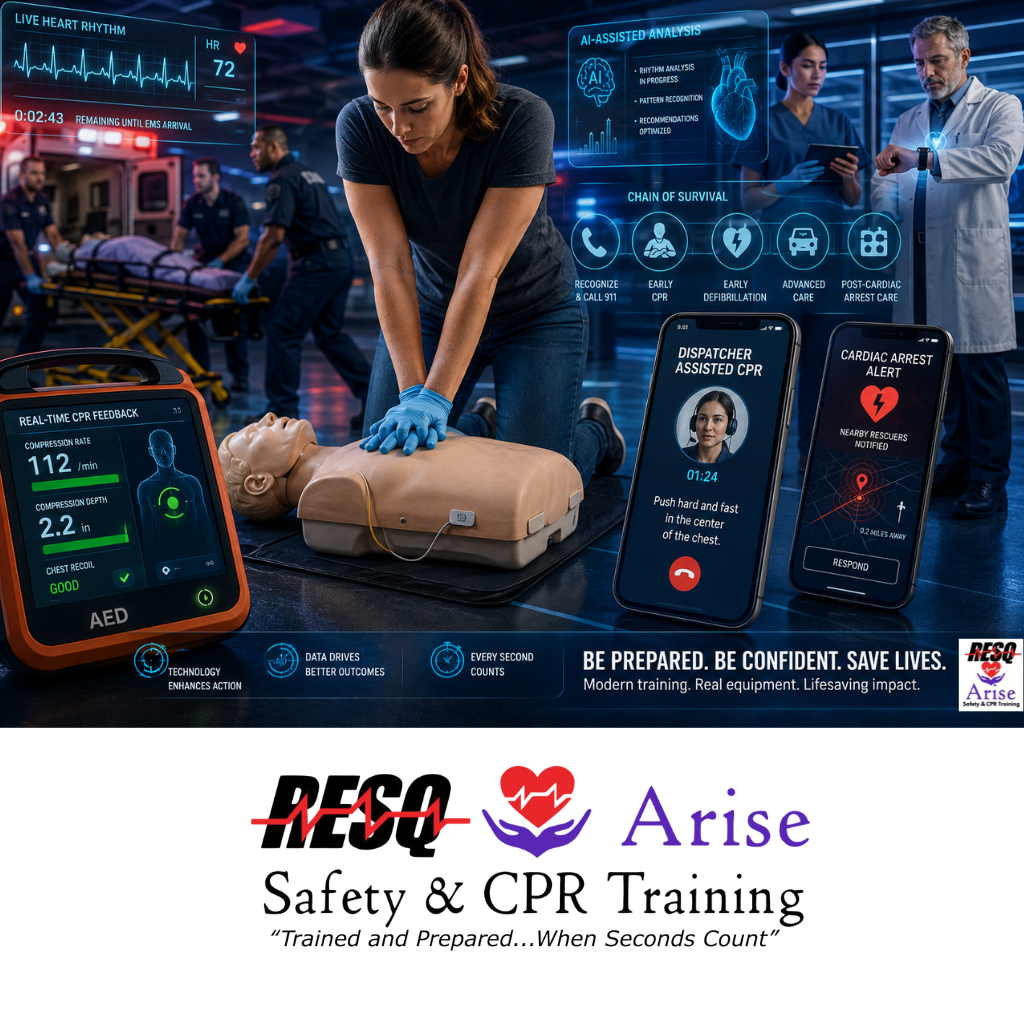
Technology has further transformed how CPR is delivered. Today, rescuers may be supported by:

  • AEDs with real-time feedback, correcting compression depth and rate as you work.
  • Smartphone apps that alert nearby trained responders to cardiac arrests in public places.
  • Dispatcher-assisted CPR, where 911 operators coach callers step-by-step through the process.
  • Wearable devices and AI systems that can detect cardiac events earlier than ever before.

Despite these advancements, one constant remains: the first few minutes still belong to the bystander. EMS response times, even in optimized systems, cannot compete with immediate action from someone already on scene. That is why modern CPR training focuses not only on technique, but also on confidence, speed, and decision-making under pressure.

In many ways, CPR has come full circle. While the tools and science have advanced dramatically, the core principle is unchanged: ordinary people stepping in to help another person in their most critical moment. From 18th-century resuscitation attempts using bellows, to today’s data-driven protocols and smart defibrillators, the mission has remained consistent—preserve life, protect the brain, and restore the heartbeat.

Learning CPR today means stepping into that legacy with the best tools and knowledge available.

Your training should reflect the modern standard.

Stay current with the latest guidelines from the American Heart Association and the American Red Cross. Get hands-on, practice with real equipment, and build the confidence to act when it matters most. Secure your spot in an upcoming class at ResqTraining.com.

Sources for Part 6:

  • Sayre, M. R., et al. (2008). Hands-Only (compression-only) cardiopulmonary resuscitation. Circulation.
  • International Liaison Committee on Resuscitation (ILCOR). 2020 International Consensus on CPR.
  • American Heart Association. High-Quality CPR Data & Guidelines (2020).
  • American Red Cross. Modern CPR Training and Accessibility Guidelines.
Part 5: The Rise of the AED

Even with high-quality CPR, there are times when the heart cannot restart on its own. That is because many sudden cardiac arrests are caused by dangerous electrical rhythms like ventricular fibrillation (VF) or pulseless ventricular tachycardia (VT). In these cases, the heart is not “stopped” in the traditional sense—it is quivering or beating so chaotically that it cannot pump blood effectively. No amount of chest compressions alone can correct this electrical problem. That is where defibrillation becomes critical.

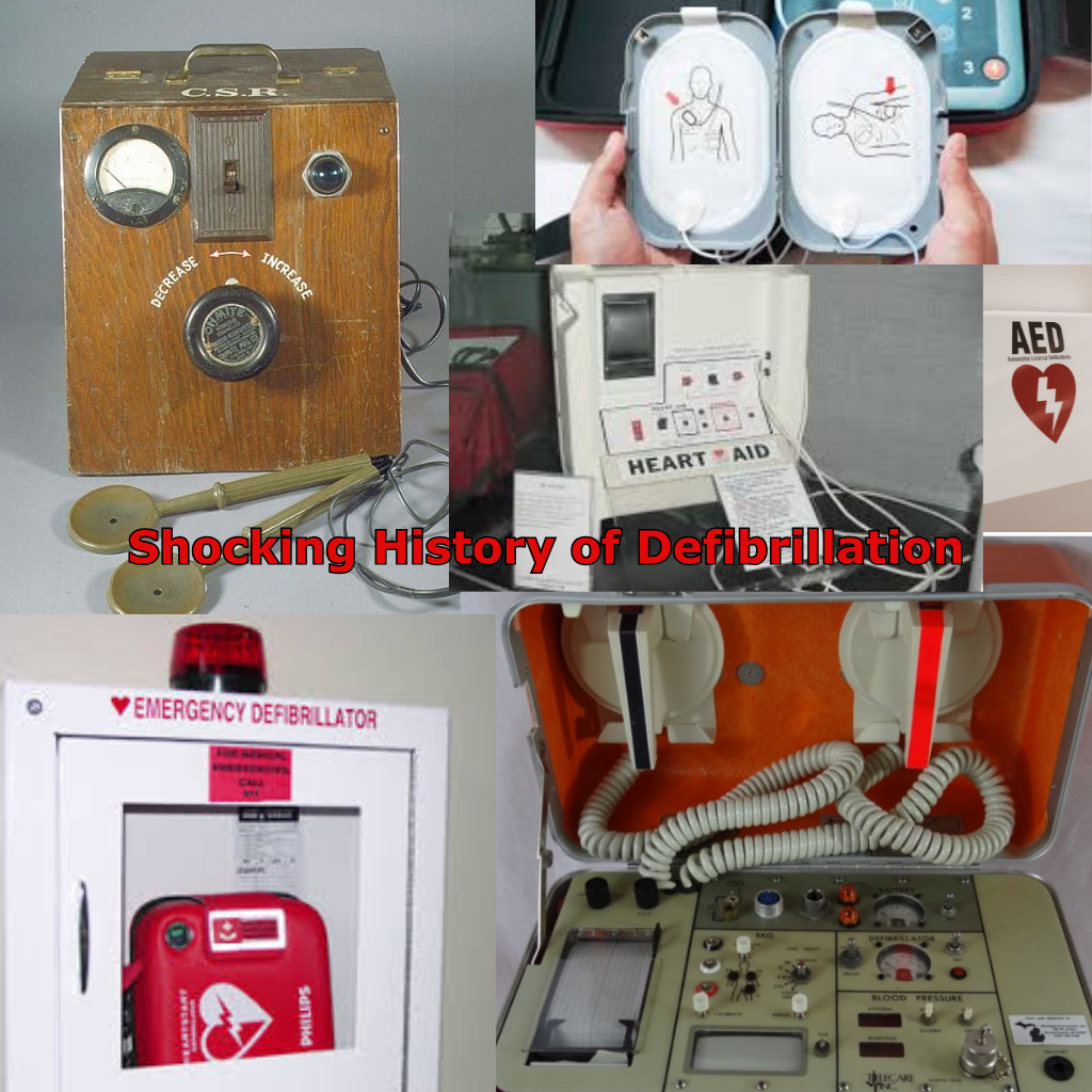
The Shocking History of Defibrillation

The concept of using electricity to restart a heart sounds like science fiction, but its roots go back to the 1700s. Early scientists observed that electricity could cause muscles to twitch. In 1775, Abildgaard proved that he could both stop a hen’s heart with an electric shock and then restart it with another. However, it wasn’t until 1947 that Claude Beck, a pioneering heart surgeon, successfully performed the first human defibrillation during surgery. At the time, this required opening the patient’s chest and applying metal paddles directly to the heart muscle.

From 100-Pound Machines to Portable Saves

Early defibrillators were anything but practical. In the 1950s and 1960s, these machines were bulky, complex, and confined to hospitals. They often weighed over 100 pounds and were powered by huge capacitors that required a wall outlet.

The real revolution began in 1965 in Belfast, Northern Ireland. Professor Frank Pantridge, often called the “Father of Emergency Medicine,” realized that if people were dying of cardiac arrest in the streets, the defibrillator needed to go to the streets. He invented the first portable defibrillator. It was still heavy—weighing 110 pounds and powered by car batteries—but it could be carried in an ambulance. This was the birth of the “Mobile Coronary Care Unit.”

The “Automated” Revolution

A major turning point came with the development of the automated external defibrillator (AED) in the late 1970s. Early inventors like Archie W. Diack helped design devices that could do something revolutionary: analyze heart rhythms automatically.

Before the AED, only doctors could decide when to shock a patient. Diack’s invention removed the need for expert interpretation by using microprocessors to “read” the heart’s electrical signals. Over time, advances in battery technology and electrode design allowed AEDs to become smaller, faster, and far more user-friendly.

How Modern AEDs Work With You

Today, AEDs are engineered specifically for the general public. When powered on, they provide clear, step-by-step voice prompts that guide the rescuer through the entire process:

  • “Apply pads to the patient’s bare chest”
  • “Analyzing heart rhythm—do not touch the patient”
  • “Shock advised—stand clear”

The device will only deliver a shock if it detects a shockable rhythm, which makes it extremely safe. You cannot accidentally shock someone who does not need it. This automation is what allows teachers, coworkers, parents, and bystanders—with little or no medical background—to intervene effectively.

The Mechanical and Electrical Partnership

AEDs and CPR are designed to work together as a coordinated system:

  • CPR keeps oxygenated blood circulating to the brain and vital organs (mechanical support).
  • AED resets the heart’s electrical system (electrical correction).

This combination dramatically increases survival rates. In fact, for every minute that defibrillation is delayed, the chance of survival decreases by about 7–10%. When CPR is started immediately and an AED is used quickly, survival rates can double or even triple. Because of this, AEDs are now commonly found in workplaces, schools, gyms, and airports.

Is there an AED in your workplace?

Knowing where it is is step one. Confidence is what saves lives. Learn how to act quickly and effectively with hands-on training at ResqTraining.com.

Sources for Part 5:

  • Pantridge, J. F., & Geddes, J. S. (1967). A mobile intensive-care unit in the management of myocardial infarction. The Lancet.
  • Diack, A. W., et al. (1979). An automatic cardiac resuscitator for emergency use. Medical Instrumentation.
  • Myerburg, R. J. (2003). The role of AEDs in public access defibrillation. Circulation.
  • Beck, C. S., et al. (1947). Ventricular fibrillation of long duration abolished by electric shock. JAMA.

Part 4: CPR Goes Public — The Rise of the Everyday Rescuer

At first, CPR was a skill reserved almost exclusively for medical professionals. Doctors, nurses, and trained hospital staff were the only ones taught how to respond to cardiac arrest. In the early 1960s, this made sense, CPR was new, and the medical community was still refining the technique.

However, a critical problem quickly became clear: most cardiac arrests weren’t happening in hospitals. They were happening at home, at work, and in public spaces, far from immediate medical care. By the time emergency responders arrived, it was often too late. Leaders in emergency medicine and organizations like the American Heart Association recognized a simple but powerful truth: survival depended on what happened in the first few minutes and that meant ordinary people needed to act.

The 1970s Shift: Training the Public & The Role of OSHA

In the 1970s, a major shift began. The American Heart Association launched large-scale efforts to teach CPR to the general public. Their goal was ambitious: turn bystanders into immediate responders.

Standardizing this shift was the arrival of the Occupational Safety and Health Administration (OSHA), which was established in 1970 and officially opened in 1971. OSHA’s mission was to ensure safe and healthful workplaces, and they quickly recognized that “safety” included emergency readiness. They began requiring that if a workplace is not in “near proximity” to a medical facility, a person must be trained to render first aid—a mandate that today strongly includes CPR and AED usage.

During this same period, training tools evolved. One of the most important innovations was the introduction of the Resusci Anne manikin. This lifelike model allowed students to physically practice chest compressions and rescue breaths in a safe, controlled environment—transforming CPR from a theoretical concept into a hands-on skill.

CPR Enters Everyday Life

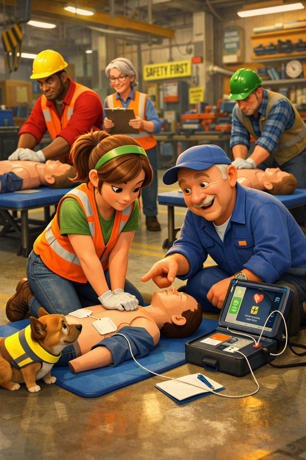
As training became more accessible and OSHA regulations moved through industries, CPR quickly spread beyond hospitals and into communities. Schools began incorporating CPR education into their programs. Workplaces started requiring employee certification for safety compliance to meet “near proximity” response time guidelines (usually interpreted as a 3-4 minute window).

For the first time in history, a regular person, without any medical background, could step in and save a life. This shift fundamentally changed the chain of survival. A neighbor, coworker, teacher, or even a stranger could now recognize cardiac arrest, begin CPR immediately, and keep blood flowing to the brain until help arrived.

A Legacy That Continues Today

What started in the 1970s has grown into a global movement. Today, millions of people are trained in CPR, and public access defibrillators (AEDs) are placed in schools, airports, gyms, and workplaces. But the mission remains the same: empower everyday people to act when seconds count. Because in a cardiac emergency, the difference between life and death often isn’t a doctor—it’s the person standing nearby who is willing and prepared to help.

Don’t Just Be a Bystander—Be a Lifesaver

You don’t need a medical degree to save a life. You just need the training—and the confidence to act. Join the millions of everyday heroes who are prepared to step in during an emergency. Sign up for a public CPR class today at ResqTraining.com and be ready when it matters most.

Sources for Part 4:

  • American Heart Association — The 1970s and the Expansion of Lay-Rescuer CPR.
  • Occupational Safety and Health Administration (OSHA). Standard 1910.151 – Medical Services and First Aid.
  • Eisenberg, M. S. (2013). Life in the Balance: A History of Combatting Sudden Cardiac Death.
  • Journal of the American College of Cardiology — The Evolution of Bystander CPR.

Part 3: Adding the Chest Compressions

Rescue breathing was a major breakthrough—but it only solved half the problem of cardiac arrest survival. Oxygen could enter the lungs, but without blood circulation, it never reached the brain or vital organs. Within minutes of a heart stopping, the lack of blood flow leads to irreversible brain damage. The missing link wasn’t just air—it was movement.

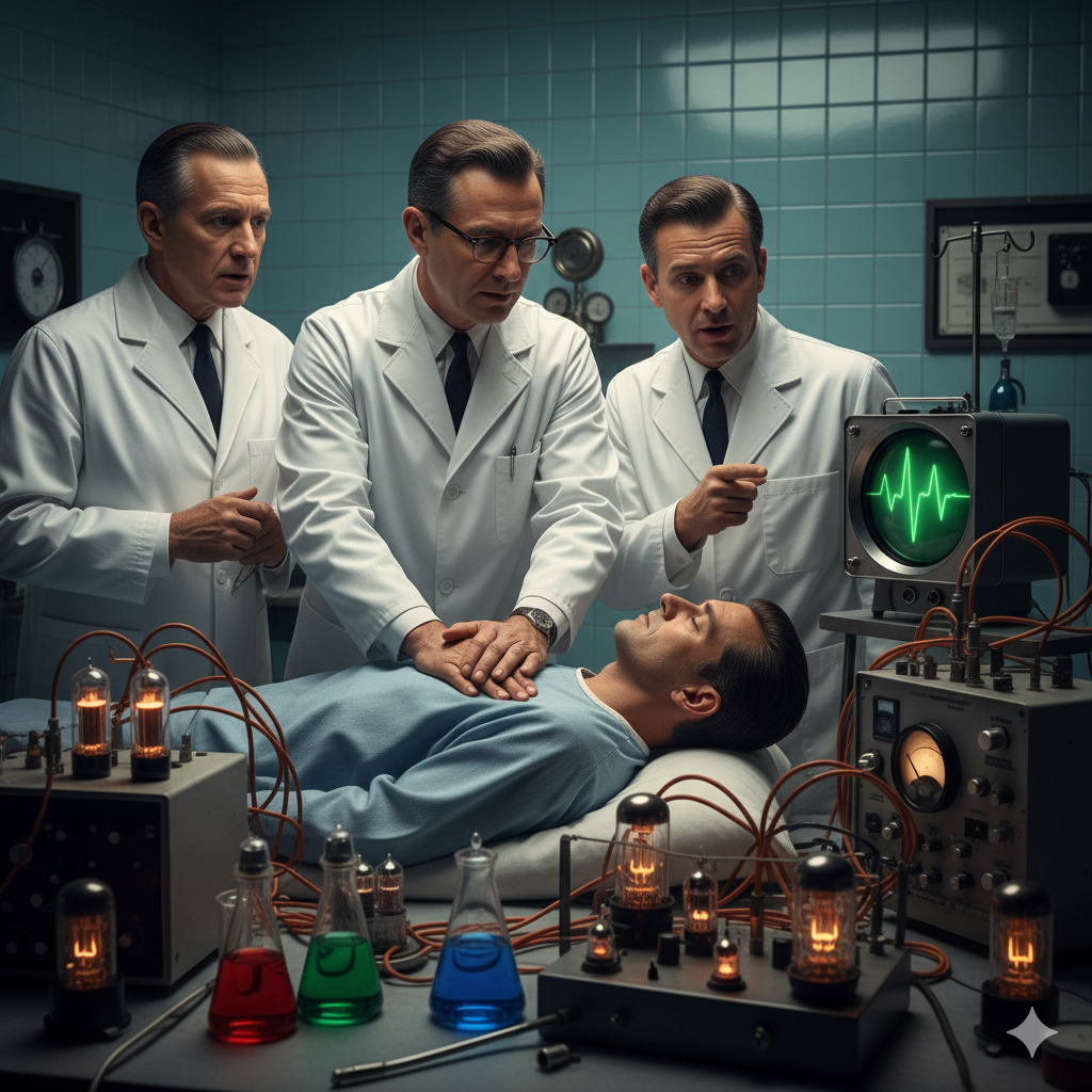
That changed in 1960 at Johns Hopkins University, when researchers William B. Kouwenhoven, James R. Jude, and Guy Knickerbocker made a discovery that would redefine emergency medicine and the history of cardiopulmonary resuscitation (CPR).

The Breakthrough: Pumping the Heart Without Surgery

While studying external electrical defibrillation—searching for a way to treat chaotic heart rhythms without invasive surgery—the team noticed something unexpected: Firm pressure applied to the chest could generate a measurable pulse. They realized that the heart could be compressed between two rigid structures:

  1. The sternum (breastbone) in the front
  2. The spine in the back

By pressing rhythmically on the sternum, blood was forced out of the heart to the brain and body. Releasing pressure allowed the heart to refill. This technique, known as “closed-chest cardiac massage,” allowed rescuers to circulate blood without opening the chest cavity. This was a massive leap from earlier invasive medical methods such as:

  • Open-Chest Cardiac Massage: A surgical procedure requiring a doctor to cut open the chest to manually squeeze the heart.
  • Internal Defibrillation: Applying shocks directly to the heart muscle during an operation.
Why Circulation Changed Cardiac Arrest Outcomes

Before this discovery, resuscitation focused almost entirely on ventilation (breathing), and survival rates remained extremely low. Afterward, rescuers could artificially create a heartbeat. This wasn’t just an improvement—it was the foundation of modern life support. Oxygen from rescue breaths could finally reach the brain, preventing cell death.

The Integration of Modern CPR Standards

When chest compressions were combined with rescue breathing, the integration formed what we now know as Cardiopulmonary Resuscitation (CPR). This coordinated system addresses both the lungs (Ventilation) and the heart (Circulation). Organizations like the American Heart Association (AHA) quickly recognized its impact and began standardizing CPR training, laying the foundation for modern emergency response protocols used worldwide today.

The Clinical Reality of High-Quality CPR

Effective chest compressions are the core of cardiac arrest intervention, but they must meet specific clinical standards to be effective:

  • Compression Depth: At least 2 inches in adults to adequately squeeze the heart.
  • Compression Rate: 100–120 beats per minute to maintain blood pressure.
  • Minimal Interruptions: To ensure constant blood flow to the brain.

In a medical emergency, circulation is the top priority, as brain damage begins in as little as 4–6 minutes. This discovery shifted the focus of first aid from a passive concept to an active, life-saving mechanical intervention.

Master the “Missing Piece” of Survival

High-quality chest compressions are the most critical factor in surviving a cardiac arrest. Build your confidence and learn proper hand placement with expert, hands-on CPR certification at ResqTraining.com.

Sources for Part 3:

  • Kouwenhoven, W. B., Jude, J. R., & Knickerbocker, G. G. (1960). Closed-chest cardiac massage. JAMA.
  • Johns Hopkins Medicine. The History of CPR: The Hopkins Connection.
  • American Heart Association. 1960: The birth of modern CPR.

Part 2: The Invention of Mouth-to-Mouth

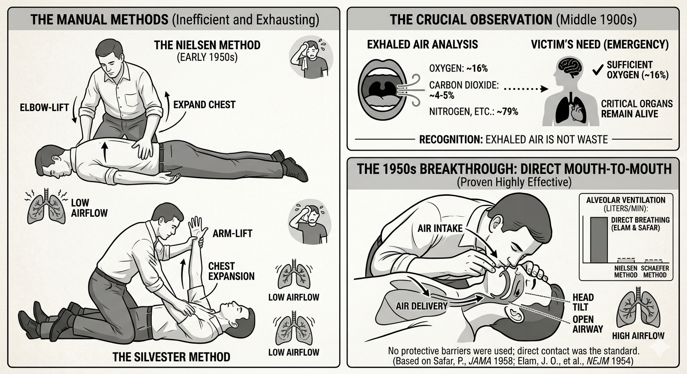
In the middle of the 1900s, scientists made a big discovery. They found that human breath still has enough oxygen to help someone else. Before this, many people believed that the air we breathed out was just waste. However, researchers discovered that while we breathe in about 21% oxygen, we only use a small amount of it. This means the air we breathe out still contains around 16% oxygen. This is more than enough to keep another person’s brain and organs alive during an emergency.

Before mouth-to-mouth became the standard, rescuers used several manual techniques that relied on body movement:

  • The Silvester Method: This involved laying the victim on their back and raising their arms above their head to expand the chest, then pressing the arms against the chest to force air out.
  • The Schaefer Method: To avoid the tongue blocking the airway, the victim was placed face-down. The rescuer would kneel over them and press on the lower back to push air out, then release to let air in.
  • The Holger Nielsen Method: Popular in the early 1950s, this combined the two. The victim was face-down, and the rescuer would pull the victim’s elbows upward to expand the chest, then press on the back to exhale.

While these were popular, they were very exhausting and moved very little air compared to direct breathing.

During the 1950s, Dr. James Elam and Dr. Peter Safar proved that direct mouth-to-mouth breathing was much more effective. They conducted experiments showing that a rescuer could maintain healthy oxygen levels in a victim just by using their own breath. Interestingly, they initially experimented with “mouth-to-nose” breathing as well, believing it might be easier to create a seal. However, they eventually settled on mouth-to-mouth as the primary method because it allowed for a larger volume of air to enter the lungs.

One major difference between then and now was the lack of protection for the rescuer. In the 1950s, there were no breathing barriers or pocket masks. Rescuers were taught to place their mouths directly onto the victim’s face. At that time, doctors were so focused on the survival of the patient that the risk of spreading germs to the rescuer was rarely discussed. It wasn’t until decades later, with a better understanding of infectious diseases, that the medical community developed the one-way valves and barriers we use today.

While these tests often focused on victims of drowning or drug overdoses, doctors realized this method could help in any situation where someone stopped breathing. Because of their hard work, this became a standard way to help victims of drowning, suffocation, and even carbon monoxide poisoning.

Consequently, doctors started to teach this method to others. It was a simple way to help, but the heart still needed more attention. Medical professionals began to notice that even if they could get air into the lungs, the victim’s skin would remain blue and their pulse would remain absent. They realized that oxygen in the lungs was useless if there was no way to transport it to the brain and other vital organs. This critical gap in knowledge meant that while rescue breathing was a massive leap forward, the “engine” of the body—the heart—was still being ignored.

Are you confident in your rescue breathing?

Learn the safest and most effective mouth-to-mouth techniques with professional guidance. Register for training at ResqTraining.com.

Sources for Part 2:

  • Safar, P. (1958). Ventilatory efficacy of mouth-to-mouth artificial respiration. JAMA.
  • Elam, J. O., et al. (1954). Oxygen and carbon dioxide exchange and alveolar ventilation in mouth-to-mask resuscitation. New England Journal of Medicine.
  • National Center for Biotechnology Information (NCBI). The history of the Holger Nielsen method.

Part 1: The Early Days of Saving Lives

In the old days, people did not understand how the heart functioned. Even without this knowledge, people tried to save those whose breathing had stopped. Although they lacked scientific understanding, they still believed that life could be restored in some cases. Because of this belief, people used their available tools and ideas to save lives.

The first techniques used to save people whose breathing had stopped were very unusual. For example, some used the “Heat Method,” which involved applying hot coals or warm ashes directly to a victim’s skin to shock the body back to life. Others tried the “Fumigation Method,” where they blew tobacco smoke into the victim’s body because they believed the warmth and nicotine would stimulate the heart. They truly believed that people could be brought back to life using these techniques. Apart from that, different groups tried various ways to restore breathing, such as rolling victims over large barrels or even hanging them upside down.

Unfortunately, these methods often caused more harm than good. The “Heat Method” frequently resulted in severe burns, while the “Fumigation Method” could cause internal damage. Over time, rescuers noticed that victims rarely woke up after these treatments. In many cases, the methods actually made the victim’s condition worse. People eventually realized these ideas did not work because the results were not consistent, and the injuries to the survivors were too great to ignore.

However, in the Middle Ages, people used even stranger techniques to save people whose breathing had stopped. For instance, people used a technique called flagellation, which meant hitting people whose breathing had stopped using whips. Another common practice was the “Inversion Method,” where victims were hung by their feet. People believed that gravity would help drain fluids and “shake” the life back into the body.

Similar to the earlier methods, these techniques were often painful and dangerous. Flagellation caused external injuries, and hanging people upside down could lead to further respiratory failure. Rescuers eventually noticed that these methods did not increase survival rates. Instead of helping, these actions often caused unnecessary suffering for the victims. Through observation, early medical thinkers realized that “waking up” the body through pain or gravity was not the answer to restoring breath.

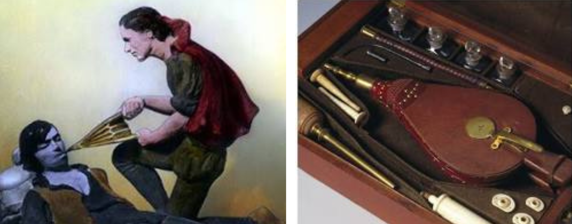
After that, new ideas began to emerge. In the 1700s, doctors began experimenting with air and breathing. For instance, doctors began using a device called a bellows. Normally, a bellows is used to blow air into a fireplace. However, doctors began using a bellows to blow air into a person’s lungs. At that time, this was a very promising idea.

In addition to bellows, doctors tried using long wooden tubes or silver pipes. They would insert these into the victim’s throat to create a direct path for air. Some societies even created a “Drowning Screen,” which was a large board used to push and pull on the victim’s chest while air was being forced in. These tools were used to manually inflate and deflate the lungs like a balloon.

Today we know that using bellows was often dangerous because the pressure could be too high for human lungs. However, this period was important for one reason. Scientists began to realize that lungs required a specific volume of air and that “stale” air was different from “fresh” air. This led to the discovery of oxygen and the understanding that breathing for someone else could keep their brain and heart alive. Driven by these early concepts, doctors began to move away from painful “shocks” and toward the scientific study of the respiratory system.

This era was the true beginning of rescue breathing. While it was only one half of the puzzle, it was a vital discovery. These early attempts at artificial ventilation paved the way for the development of full CPR, which would eventually save millions of lives.

Sources:

  • The American Heart Association: History of CPR
  • Museum of Bernoulli: Early Respiratory Devices
  • Journal of Emergency Medicine: Evolution of Resuscitation

RESQ Arise | CPR, First Aid & BLS Training in Central Indiana